Where Gliomas Start — And Why Location Should Shape Surgery
Minimal brain graphic highlighting the brainstem; title ‘Where Gliomas Start — Brainstem.
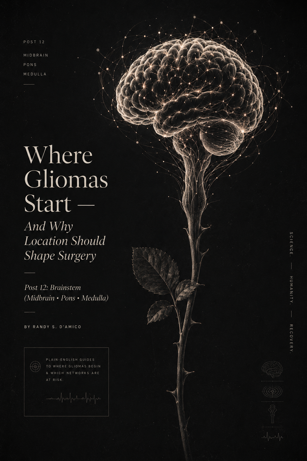
Post 12: Brainstem (Midbrain • Pons • Medulla)
About this series. Plain-English guides to where adult gliomas begin and which brain networks are at risk—so patients and families understand planning, mapping, and recovery.
Why the brainstem is different
The brainstem is the narrow stalk connecting brain to spinal cord. It’s small, dense, and essential. In a few centimeters it contains:
the main motor and sensory highways for the whole body
the control centers for eye movements, facial function, swallowing, and voice
circuits involved in breathing, heart rate, and alertness
That’s why in the brainstem, millimeters matter. Even benign-appearing lesions can cause major symptoms, and treatment decisions are often less about “removing everything” and more about protecting function.
Takeaway: Brainstem tumors are about pathways and safety strategy first—then tumor control.
The brainstem map
Think of three stacked levels:
1) Midbrain (top)
Main jobs: eye movement control, pupil function, arousal circuits
Common cranial nerves here: III (oculomotor), IV (trochlear)
If affected: double vision, eyelid droop, pupil changes, imbalance
2) Pons (middle)
Main jobs: face sensation/movement, hearing/balance integration, coordination links
Cranial nerves here: V (trigeminal), VI (abducens), VII (facial), VIII (vestibulocochlear)
If affected: facial numbness/weakness, eye movement problems, vertigo, hearing symptoms, ataxia
3) Medulla (bottom)
Main jobs: swallowing, voice, breathing and heart regulation
Cranial nerves here: IX (glossopharyngeal), X (vagus), XI (accessory), XII (hypoglossal)
If affected: dysphagia, hoarseness, weak cough, tongue weakness, breathing issues
The “long tracts” (the major highways passing through)
These are the most important “wires” to picture:
Corticospinal tract — strength and motor control
What it does: carries “move” signals from brain to spinal cord
If affected: weakness, clumsiness, spasticity, slowed fine motor
Medial lemniscus — fine touch and position sense
What it does: vibration/position sense (proprioception)
If affected: numbness, poor balance in the dark, clumsy hand placement
Spinothalamic tract — pain and temperature
What it does: pain/temp pathways
If affected: altered pain/temp sensation on the opposite side of the body
Reticular activating system — alertness
What it does: keeps you awake and cognitively “online”
If affected: unusual sleepiness, slowed processing, fluctuating attention
Common signs you might notice
Brainstem symptoms often come in combinations because everything is tightly packed:
Double vision or trouble moving one eye (especially looking sideways)
Facial numbness or tingling, sometimes with jaw pain
Facial weakness (droop, eye closure trouble)
Unsteady gait / ataxia (especially with pons/cerebellar connections)
Swallowing trouble, coughing with liquids, hoarse voice
Weakness on one side or both
Odd sensory changes (pain/temp vs vibration/position feel different)
Sleepiness or mental “fog” that seems out of proportion
Red flags: progressive swallowing difficulty, weak cough, breathing changes, or rapid neurologic decline deserve urgent evaluation.
How teams plan care (Before • During • After)
Before treatment
Clarify the goal: In adults, many brainstem lesions are treated with a strategy of diagnosis + targeted therapy, rather than aggressive resection.
Imaging detail matters: high-quality MRI (with diffusion and contrast patterns) to estimate whether lesion is focal/exophytic vs infiltrative.
Decide on tissue diagnosis: biopsy can be essential when imaging is uncertain or when molecular information will change therapy.
Baseline exam: eye movements, facial sensation/motor, swallowing/voice, strength, coordination, and a clear symptom diary.
During procedures (when biopsy or surgery is done)
Route selection is everything: surgeons choose corridors that minimize crossing long tracts and protect perforating vessels.
Neuromonitoring: motor/sensory evoked potentials and cranial nerve monitoring when relevant.
A strict stopping rule: if mapping/monitoring signals risk to key pathways, the safest move is to stop.
After treatment
Swallow and airway first (medulla/lower pons): early speech-language evaluation, diet modification, cough-strength strategies.
Eye/vision rehab: prisms, patching strategies, vestibular therapy when dizziness is prominent.
PT/OT: gait stability, fall prevention, and fine motor retraining.
Fatigue/alertness plan: sleep optimization, pacing, and cognitive rehab when needed.
Ask your surgeon (patient-facing)
Which brainstem level is involved—midbrain, pons, or medulla—and what does that predict?
Do you recommend biopsy to guide therapy, and what route would you use?
What neuromonitoring will be used to protect motor/sensory tracts and cranial nerves?
What symptoms should trigger urgent reassessment (swallowing, breathing, vision)?
Bottom line
Brainstem tumors sit beside the nervous system’s most essential pathways. Good care is strategy-driven: define the goal, get the right diagnosis, choose corridors carefully, and protect long tracts and cranial nerves. Recovery often depends as much on early rehab—swallowing, eye control, gait—as it does on the tumor itself.
Fast FAQ
Are brainstem tumors always inoperable?
No. Some are focal or grow outward (exophytic) and may be partially resectable. Many adult brainstem tumors, however, are best managed with biopsy plus medical therapy.
Why is biopsy sometimes recommended even when MRI “suggests” a diagnosis?
Because treatment increasingly depends on molecular features, and imaging alone can be misleading.
Can symptoms improve?
Often yes—especially if symptoms are driven by swelling or compression. Rehab is critical for swallowing, gait, and eye control.