Where Gliomas Start — And Why Location Should Shape Surgery
Cerebellar tumors in plain English—vermis vs hemisphere vs flocculonodular anatomy, dentate/SCP pathways, hydrocephalus red flags, and recovery.
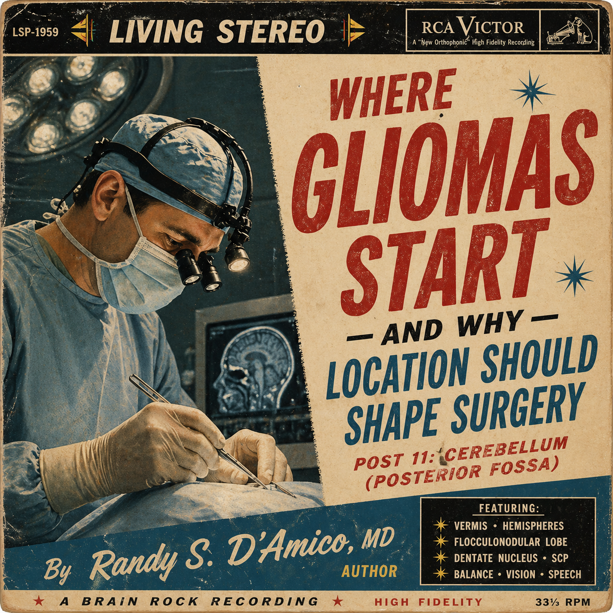
Post 11: Cerebellum (Posterior Fossa)
About this series. Plain-English guides to where adult gliomas begin and which brain networks are at risk—so patients and families understand planning, mapping, and recovery.
Why the cerebellum matters
The cerebellum sits in the back of the head, below the cerebral hemispheres. Most people associate it with balance. But it’s better described as the brain’s timing and calibration engine—for movement, eye control, speech rhythm, and even the “smoothness” of thinking.
The posterior fossa is also a tight space. Small changes from swelling or CSF blockage can cause symptoms quickly. So planning is about networks and pressure/CSF pathways.
Takeaway: Cerebellar tumors can affect walking, eye movements, coordination, and speech cadence. Location inside the cerebellum predicts which of those are most at risk.
Cerebellar anatomy in one minute (the map that matters)
Think of the cerebellum in three layers:
1) Vermis (midline)
Function: trunk posture, gait stability, “staying upright.”
Symptoms if affected: wide-based walking, veering, falls on turns.
2) Cerebellar hemispheres
Function: fine control of limbs—precision reaching, smooth hand movements.
Symptoms if affected: overshooting targets (dysmetria), tremor-like wobble, clumsy hands.
3) Flocculonodular lobe (inferior/posterior, near brainstem)
Function: balance integration and eye stabilization (vestibular system).
Symptoms if affected: vertigo-like dizziness, nystagmus (eye jitter), nausea, “bouncing” vision.
And the key output “highways” that carry cerebellar signals:
Dentate nucleus (deep cerebellar “output station” for coordination and cognitive pacing)
Superior cerebellar peduncle (SCP) → crosses to the other side → red nucleus/thalamus → motor and prefrontal cortex
Middle cerebellar peduncle (MCP) brings in cortical information via the pons (planning inputs)
Inferior cerebellar peduncle (ICP) brings vestibular and sensory inputs (balance and body-state)
Cerebellar networks in plain English (with anatomy)
1) Cerebello–thalamo–cortical loop— timing & smoothness circuit
Anatomy: dentate nucleus → SCP → (crosses) → thalamus → motor/premotor and prefrontal cortex.
What it does: smoothness of movement, motor learning, pacing; also contributes to “mental tempo.”
If affected: clumsiness, slowed coordination; sometimes “my thinking feels delayed,” especially when dentate/SCP are involved.
2) Vestibulo-ocular network— balance + gaze stability
Anatomy: flocculonodular lobe + vestibular nuclei ↔ ICP pathways; integrates with eye movement centers.
What it does: keeps vision stable when your head moves (VOR) and keeps the world from “bouncing.”
If affected: dizziness, nausea, nystagmus, double vision, motion intolerance.
3) Spinocerebellar / proprioceptive calibration— where your body is in space
Anatomy: sensory/proprioceptive inputs via ICP, plus midline vermis processing.
What it does: calibrates posture and gait in real time.
If affected: unsteady stance, “drunk walk” feeling, worsens in low light.
4) Cerebellar speech timing— rhythm and cadence
Anatomy: hemispheric cerebellum + dentate outputs to speech-motor planning cortex.
What it does: makes speech smooth and rhythmic.
If affected: scanning speech (choppy cadence), slurring, fatigue-related speech breakdown.
Common signs you might notice
Gait issues (vermis): wide stance, veering, trouble turning, falls.
Limb coordination (hemisphere): overshooting when reaching, shaky handwriting, dropping objects.
Eye/balance (flocculonodular/ICP): dizziness, nausea, bouncing vision, double vision.
Speech (dentate/SCP): choppy rhythm, slurred articulation.
Pressure red flags: headache + vomiting + increasing sleepiness can signal CSF blockage.
The neighbors that shape urgency
Brainstem: vital pathways + cranial nerve nuclei (swallowing, facial movement, eye control).
Fourth ventricle / aqueduct: CSF flow channels—blockage can cause hydrocephalus.
Cranial nerves and vascular structures: delicate in the posterior fossa; traction matters.
How teams plan surgery (Before • During • After)
Before surgery
Define location precisely: vermis vs hemisphere vs flocculonodular region; relationship to dentate and SCP.
Hydrocephalus plan: evaluate fourth ventricle compression and decide if CSF diversion is needed.
Baseline exam: gait, finger-to-nose, heel-to-shin, eye movements (nystagmus), swallowing screen if needed.
During surgery
Corridor selection: midline vs hemispheric approach based on which cerebellar “zone” is involved.
Monitoring: brainstem/cranial nerve monitoring when close; careful handling of cerebellar tissue to avoid traction injury.
Protect outputs: be deliberate around the dentate nucleus and SCP when they’re near the tumor—these are high-yield pathways for long-term coordination.
After surgery
Early mobilization: gait and balance improve with practice—PT begins early.
Vestibular rehab: gaze stabilization exercises, graded motion exposure, nausea control.
Speech therapy: for scanning speech or slurring.
Watch pressure: worsening headache/vomiting/somnolence warrants urgent reassessment.
Ask your surgeon (patient-facing)
Is the tumor closer to the vermis, a hemisphere, or the flocculonodular region?
Are the dentate nucleus or superior cerebellar peduncle nearby?
What’s my risk of hydrocephalus, and how will you monitor/treat it?
What rehab starts right away (balance, vestibular, speech)?
Bottom line
Cerebellar tumors affect networks for coordination, balance, gaze stability, and speech timing. Anatomy predicts function: vermis = gait, hemisphere = limbs, flocculonodular = dizziness/eyes, dentate/SCP = output and pacing. With careful corridors, pressure management, and early rehab, many patients recover stability and independence.
Fast FAQ
Is dizziness from a cerebellar tumor the same as an inner ear problem?
It can feel similar, but here the problem is the brain’s integration of balance and eye signals—not the ear alone.
Can balance improve after surgery?
Often yes—especially when swelling resolves and vestibular/gait therapy starts early.
Why are headaches and vomiting such a big deal in posterior fossa tumors?
They can signal rising pressure from blocked CSF flow. That pattern deserves urgent evaluation.