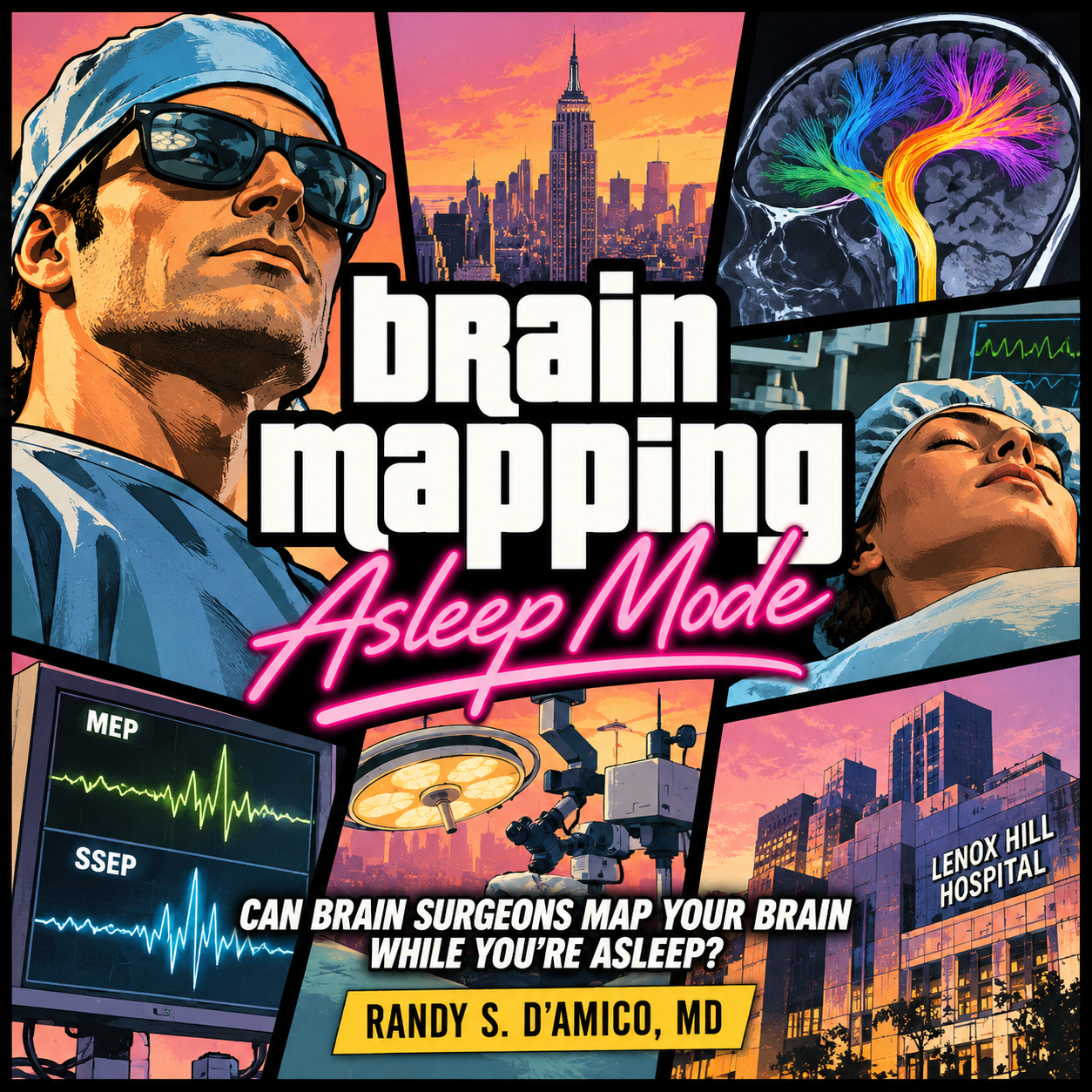
Your brain doesn't have to be awake for us to communicate with it. During brain tumor surgery, we can use electrical stimulation to identify critical pathways and monitor the signals controlling movement—even while you're under anesthesia. The mission? Remove the tumor. Protect the person.
How electrical stimulation, real-time monitoring, and modern imaging help protect movement during brain tumor surgery.
Imagine having a brain tumor removed while you're completely asleep.
You cannot speak, move your hands, or tell your surgeon whether something feels different. So how can the surgical team know whether the operation is approaching an area of the brain responsible for movement?
The answer is that your nervous system can communicate even when you're unconscious.
Using electrical stimulation and sophisticated monitoring equipment, surgeons can test specific pathways in the brain and spinal cord during an operation. These techniques help identify critical structures, monitor their function, and guide decisions about how much tumor can safely be removed.
This is called asleep brain mapping, and it is an important part of modern brain tumor surgery.
Your Brain Is an Electrical Network
Every time you move your hand, wiggle your toes, or take a step, electrical signals travel through a complex network of neurons.
The process begins in regions of the brain involved in planning and initiating movement. Signals travel through the motor cortex, descend along white matter pathways, pass through the brainstem, and ultimately reach the spinal cord and muscles.
One of the most important pathways is the corticospinal tract.
Think of it as a major communication highway connecting the brain to the body.
Damage to this pathway can cause weakness or paralysis, even when the part of the brain responsible for initiating movement remains intact.
This distinction is especially important in brain tumor surgery.
A tumor may be located far from the surface of the brain, yet still lie immediately adjacent to the white matter pathways that control movement.
Protecting these connections can be just as important as protecting the brain's outer surface.
How Do Surgeons Test Movement When You're Asleep?
During surgery, specialized electrodes can stimulate parts of the nervous system while other electrodes record the resulting electrical activity.
Two techniques are particularly important.
1. Motor Evoked Potentials: Monitoring the Movement Pathways
Motor evoked potentials, or MEPs, help surgeons assess whether signals can travel from the brain to the muscles.
Electrical stimulation is delivered to the motor system, and electrodes placed in muscles record the resulting responses.
If those responses change significantly during surgery, the team investigates whether the motor pathways may be under stress.
This can happen because of mechanical manipulation, changes in blood supply, or other physiological factors.
Importantly, a change in an MEP does not automatically mean permanent injury has occurred.
It is a warning signal that must be interpreted in context.
The surgical team may pause, adjust its technique, optimize physiological conditions, or reassess the operative strategy.
The goal is to identify potentially reversible problems before they become lasting neurological deficits.
2. Direct Electrical Stimulation: Finding the Boundaries
Monitoring tells us whether a pathway is functioning.
Direct electrical stimulation helps us identify where that pathway is located.
During surgery, a surgeon can apply small electrical currents directly to brain tissue using a specialized probe.
When stimulation activates motor-related structures, the team may detect a response in the patient's muscles.
This allows the surgeon to identify functional tissue and pathways even when the patient is under general anesthesia.
For example, while removing a tumor deep within the frontal lobe, stimulation of tissue near the surgical cavity may produce a response in the hand or leg.
That information helps the surgeon understand the relationship between the tumor and the patient's motor pathways.
As the operation progresses, repeated stimulation can help define the functional boundaries of the resection.
The exact interpretation depends on the stimulation technique, anesthesia, anatomy, and other factors. Electrical responses are not a perfect measurement of distance or safety.
But they provide valuable information that anatomical imaging alone cannot.
What About Sensation?
Movement is only one part of neurological function.
The brain also receives a continuous stream of sensory information from the body.
Surgeons can monitor some of these pathways using somatosensory evoked potentials, or SSEPs.
Small electrical impulses are delivered to peripheral nerves, commonly at the wrist or ankle. The resulting signals are recorded as they travel through the nervous system.
Changes in these responses may indicate that sensory pathways are being affected.
SSEPs can also help identify the central sulcus, an important anatomical boundary separating the primary motor and sensory cortices.
Together, MEPs, SSEPs, and direct electrical stimulation provide complementary information.
One technique cannot replace all the others.
Brain Mapping Is More Than Watching a Screen
Modern brain tumor surgery often begins long before the patient enters the operating room.
Preoperative MRI can identify the tumor and surrounding anatomy.
Diffusion MRI tractography can estimate the location of major white matter pathways.
Functional imaging and connectomics may provide additional information about how the tumor interacts with broader brain networks.
These tools help surgeons develop a plan.
But there is an important limitation.
A brain map created before surgery is not the same as a functional measurement obtained during surgery.
As tissue is removed, the brain can shift. The relationship between the tumor and surrounding structures can change.
And tractography, while valuable, is a computational estimate rather than a direct demonstration of whether a pathway is functioning.
This is why intraoperative mapping remains important.
Preoperative imaging provides the roadmap. Intraoperative stimulation and monitoring help test that roadmap against the patient's actual physiology.
Does Everyone Need to Be Awake for Brain Tumor Surgery?
No!
Awake brain surgery can be particularly valuable when surgeons need to test complex functions such as speech, language, or certain cognitive abilities.
These functions often require the patient to actively participate in testing.
But many motor pathways can be assessed under general anesthesia.
For some tumors, asleep mapping provides the information needed to guide a safe resection without requiring the patient to be awake.
In other situations, an awake procedure may offer important advantages.
The decision depends on several factors, including the tumor's location, the neurological functions at risk, the patient's health and preferences, and the capabilities of the surgical team.
Neither approach is universally superior.
The objective is not to perform the most technologically elaborate operation. It is to choose the mapping strategy that best protects the functions that matter to that patient.
Can Mapping Prevent Every Neurological Deficit?
Unfortunately, no.
Brain tumors can distort normal anatomy, infiltrate functioning tissue, and affect the blood vessels supplying critical regions.
Even with advanced monitoring, some injuries cannot be predicted or prevented.
Electrical signals can also remain stable despite certain types of neurological injury.
Furthermore, monitoring motor pathways does not necessarily tell us whether language, memory, attention, or other complex functions will be preserved.
This is one reason modern brain tumor surgery increasingly combines multiple sources of information.
The surgeon must interpret the anatomy, the imaging, the patient's baseline neurological function, the intraoperative findings, and the goals of treatment.
Sometimes the safest decision is to leave a small amount of tumor behind.
That is not necessarily a failure of surgery.
It may represent the best balance between removing disease and preserving the patient's independence and quality of life.
Questions to Ask Your Brain Tumor Surgeon
If you or someone you love is preparing for brain tumor surgery, consider asking:
Is my tumor near a pathway responsible for movement, sensation, or language?
Will you use electrical stimulation or neurological monitoring during surgery?
Would awake or asleep mapping be more appropriate for my tumor?
How will you determine when further tumor removal becomes too risky?
What neurological changes are possible after surgery, and which may improve with rehabilitation?
These conversations are especially important when a tumor is located near critical functional structures.
The Bottom Line
Brain tumor surgery is not simply about removing abnormal tissue.
It is about understanding which parts of the brain and its connections must continue working after the operation.
Asleep brain mapping allows surgeons to gather functional information from the nervous system while the patient remains under general anesthesia.
Combined with modern imaging, direct electrical stimulation, and careful surgical judgment, these techniques can help guide safer and more individualized operations.
The goal is not merely to remove as much tumor as possible.
It is to remove as much tumor as safely possible while preserving the neurological functions that make everyday life possible.
Frequently Asked Questions
Can surgeons test my brain while I'm unconscious?
Yes. Electrical stimulation and neurophysiological monitoring can assess certain motor and sensory pathways during general anesthesia.
Does brain mapping hurt?
During asleep mapping, the patient is under anesthesia and does not consciously experience the stimulation. Muscle responses can still occur.
Is awake brain surgery always better?
No. Awake and asleep mapping serve different purposes. The appropriate technique depends on the tumor's location and the neurological functions that need to be tested.
Can brain mapping guarantee that I won't have weakness after surgery?
No. Mapping can help identify and monitor important pathways, but it cannot eliminate every risk of neurological injury.
What is the difference between tractography and electrical stimulation?
Tractography estimates the location of white matter pathways using MRI data. Electrical stimulation tests physiological responses during surgery. They provide complementary information.
