How Do Brain Surgeons Know What’s Safe to Remove?
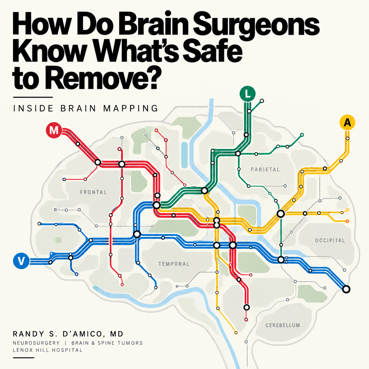
Inside Brain Mapping During Brain Tumor Surgery
One of the most important questions in brain tumor surgery is also one of the simplest:
How does the surgeon know where to stop?
Brain mapping is a technique used during brain surgery to identify areas and pathways responsible for important functions such as movement, speech and language, helping surgeons determine what tissue can be safely removed.
MRI can show us where a tumor is. It can show us how big it is. It can show us which structures are nearby.
But MRI alone cannot always tell us exactly what a specific piece of brain tissue is doing in a specific person. That is where brain mapping comes in.
Brain mapping allows us to test brain function during surgery and identify the regions and pathways that matter most for movement, speech, language, vision, attention, and other abilities.
In simple terms: Imaging helps us predict where function may be. Mapping helps us test it.
That distinction can shape how much tumor we can safely remove.
Believe it or not! The brain does not come with labels!
If you open a textbook, the brain looks beautifully organized. Language here. Movement there. Vision in the back. Memory in the temporal lobe.
Real brains are slightly more complicated.
There is enormous consistency in how the nervous system is organized, but there is also meaningful variation from person to person.
Tumors make that even more complicated. A slow-growing tumor can distort anatomy over years. Nearby regions may adapt. Networks may reorganize. White-matter pathways may be displaced rather than destroyed.
So even when we have excellent imaging, the most important question is often not:
“Where should this function be?”
It is:
“Where is this function in this patient?”
Brain mapping starts before the operation
The mapping process does not begin when the surgeon touches the brain. It begins during planning.
Depending on the case, that may include:
structural MRI
diffusion imaging and tractography
functional MRI
connectomic analysis
neuropsychological testing
detailed language or motor assessment
the neurological examination
These tools help us build a working map of the tumor and the networks around it. But this is still a hypothesis. The operating room is where we can sometimes test that hypothesis directly.
What is direct electrical stimulation?
During brain mapping, a surgeon can apply a very small electrical stimulus to the surface of the brain or to tissue deeper within it.
The stimulation briefly interrupts or activates the function of that area. The effect is temporary. It does not mean the tissue has been damaged. Think of it like briefly interrupting one connection in a complex electrical system to see what changes.
If a patient is naming pictures and stimulation repeatedly causes the same naming error, that location may be important for language.
If stimulation causes a hand or arm to move, we have identified part of the motor system.
If speech suddenly stops while everything else remains intact, we may have found tissue essential for speech production.
These responses help create a functional map in real time.
Mistakes during mapping are useful!
This often surprises patients. During mapping, a temporary mistake can be exactly what we are looking for.
Imagine a patient is repeatedly shown pictures: Dog, Chair, Apple, Umbrella. Then, during stimulation of one small area, the patient suddenly cannot name the umbrella. A few seconds later, stimulation stops and the word comes back.
That is not a failed test. It is valuable information. It tells us that disturbing that region affects a function we are trying to preserve. A temporary error during mapping may help prevent a permanent one after surgery.
Motor mapping
Movement is one of the clearest examples of why mapping matters.
The motor cortex sends signals down through deep white-matter pathways, including the corticospinal tract, before those signals reach the spinal cord and muscles.
During surgery, we may use electrical stimulation to identify:
the motor cortex
pathways controlling the face
pathways controlling the hand
pathways controlling the leg
deeper motor fibers beneath the tumor
We may also use neurophysiological monitoring, including motor evoked potentials and sensory signals, to watch those systems continuously. These techniques help us understand how close the operation is getting to structures that could affect strength or movement.
Language mapping is more complicated
Language is not stored in one tiny spot in the brain. It is a network.
Speaking requires multiple processes:
finding the right word
understanding meaning
producing sounds
sequencing speech
reading
repeating
connecting concepts
coordinating the muscles involved in speech
That is why language mapping may involve several different tasks. During surgery, a patient might:
name pictures
read words
repeat sentences
count
describe images
complete phrases
answer simple questions
The exact tasks depend on where the tumor is and which language networks may be at risk. A tumor near speech production pathways requires different testing than one near semantic or reading networks.
Cortical mapping versus subcortical mapping
This distinction is especially important.
Cortical mapping
This tests the brain's surface. It helps identify areas of cortex involved in functions such as movement or language. But removing a tumor is rarely only about the surface. Many gliomas extend deeper into the brain.
That is where white matter becomes critical…
Subcortical mapping
As surgery progresses deeper, stimulation can be used to test the pathways beneath the cortex. These white-matter tracts are the brain's communication cables. They connect distant regions into functioning networks.
Examples include pathways involved in:
movement
language
vision
attention
speech initiation
semantic processing
This is one of the reasons I think of modern brain surgery less as removing a mass and more as navigating a network.
Why white matter matters so much
Cortex gets much of the attention because it is easy to visualize. But white-matter pathways may be even less forgiving. A portion of cortex may sometimes be compensated for by neighboring regions. A major communication highway can be much harder to replace.
That means a surgeon may safely remove tumor through one region, only to reach a deeper boundary where the risk suddenly changes. This is why the last few millimeters of a resection can matter so much. And it is one reason direct subcortical mapping can help determine when the safest decision is to stop.
What about vision, attention, and higher-level cognition?
Brain mapping is evolving beyond traditional motor and language testing. In selected cases, teams may also test functions such as:
reading
visual processing
spatial attention
executive function
working memory
social cognition
These functions are harder to test reliably in the operating room. They often depend on widely distributed networks rather than one isolated region. That makes them one of the most interesting frontiers in functional neurosurgery. The broader lesson is that preserving function means thinking beyond whether a patient can simply move an arm or say their name.
Where does connectomics fit?
Connectomics is the study of how different parts of the brain are connected into networks.
For brain tumor surgery, connectomic imaging can help us understand which large-scale networks may run through or around a tumor. That can be incredibly useful for planning. But imaging and direct mapping serve different purposes.
Connectomics may help answer:
“Where might an important network be?”
Direct stimulation may help answer:
“Does disrupting this specific location actually affect function right now?”
The two approaches can complement each other. One helps build the roadmap. The other can test the road while we are traveling on it.
Is brain mapping always done while the patient is awake?
No. Some mapping can be performed under general anesthesia. Motor pathways, for example, can often be mapped and monitored without the patient being awake.
Language is different. If we need to know whether stimulation affects naming, reading, comprehension, or speech, we need the patient to perform those tasks. That is why certain tumors near important language networks may be approached with awake brain mapping.
The patient does not need to be awake for the entire operation. In many cases, they are asleep for portions of the procedure and awake only during the period when functional testing is needed.
Does mapping make surgery safer?
The goal of mapping is to increase the amount of useful information available during surgery. For appropriately selected tumors, particularly those near critical functional areas, direct mapping can help identify boundaries that would be difficult to determine from anatomy alone.
That can support two equally important goals:
Remove as much tumor as safely possible.
and
Preserve the neurological function that matters to the patient.
Neither goal should exist without the other.
What happens when mapping says “stop”?
This is where brain mapping connects directly to the idea of maximal safe resection. Imagine that most of a tumor has been removed. There is still abnormal tissue visible. But stimulation now repeatedly produces a language error.
Or motor stimulation shows that the corticospinal tract is extremely close. At that point, the remaining tumor may carry a very different risk than what has already been removed. That is not necessarily a failed operation. It may mean we have found the functional boundary. Good surgery is not simply about knowing how to remove tumor. Sometimes it is about knowing when not to.
Can brain mapping be wrong?
No test in medicine is perfect. Mapping results have to be interpreted in context. Fatigue can affect performance. Seizures can interfere with testing. Tumors can distort anatomy. Stimulation parameters matter. Tasks have to be selected carefully. That is why mapping is not a single button or device.
It is a process that combines:
surgical anatomy
imaging
physiology
patient performance
neuropsychology
experience
judgment
The value comes from putting those pieces together.
What should patients ask their surgeon?
If a tumor is close to an important functional area, reasonable questions include:
Which networks are closest to my tumor?
What functions are most at risk?
Will you use direct brain mapping?
Will I need to be awake for part of the surgery?
Will you use subcortical mapping as well as cortical mapping?
How will you know when it is time to stop removing tumor?
What other tools will you use to protect function?
Those questions often reveal more about the surgical strategy than simply asking how many hours the operation will take.
The bigger picture
Brain tumor surgery has traditionally been described as an anatomical problem: Find the tumor. Expose it. Remove it. But the brain is not simply anatomy. It is a living network. And increasingly, the challenge is not just seeing the tumor more clearly. It is understanding the individual brain around it more clearly.
The future of brain tumor surgery is not simply better tumor visualization. It is better understanding of the person’s functional brain.
And when we can combine imaging, network analysis, direct stimulation, and careful clinical judgment, the goal becomes more precise:
remove what can safely be removed, protect what cannot be replaced, and preserve the life the patient wants to return to.
Frequently Asked Questions
What is brain mapping during brain tumor surgery?
Brain mapping is the process of identifying brain regions and pathways involved in important functions such as movement, speech, and language. It can involve imaging before surgery and direct electrical stimulation during surgery.
Is brain mapping painful?
No. The brain itself does not contain pain receptors. During awake mapping, the scalp is numbed and the anesthesia team focuses on keeping the patient comfortable.
Why do surgeons stimulate the brain?
Electrical stimulation can temporarily alter a specific function, helping surgeons identify areas or pathways that should be preserved.
What is subcortical brain mapping?
Subcortical mapping tests the white-matter pathways beneath the brain's surface. These pathways connect different parts of the brain and may define the functional boundary of a tumor resection.
Do all brain tumor surgeries require mapping?
No. Mapping is most useful when a tumor is near brain regions or pathways where injury could significantly affect neurological function.
Can brain mapping help surgeons remove more tumor?
In selected cases, mapping can provide greater confidence about which tissue can be safely removed while identifying functional boundaries that should be preserved.
Bottom line
Brain mapping helps answer one of the hardest questions in neurosurgery:
Where does safe tumor removal end and unacceptable functional risk begin?
MRI, tractography, connectomics, monitoring, and direct stimulation each provide part of the answer.
Together, they allow brain tumor surgery to become increasingly individualized—not simply to the tumor, but to the person carrying it.
Educational content only and not a substitute for individualized medical advice.
