Where Gliomas Start — And Why Location Should Shape Surgery - SMA & Pre-SMA (Medial Frontal Lobe)
SMA & Pre-SMA: the brain’s “start signal” for movement and speech—why temporary SMA syndrome often improves.
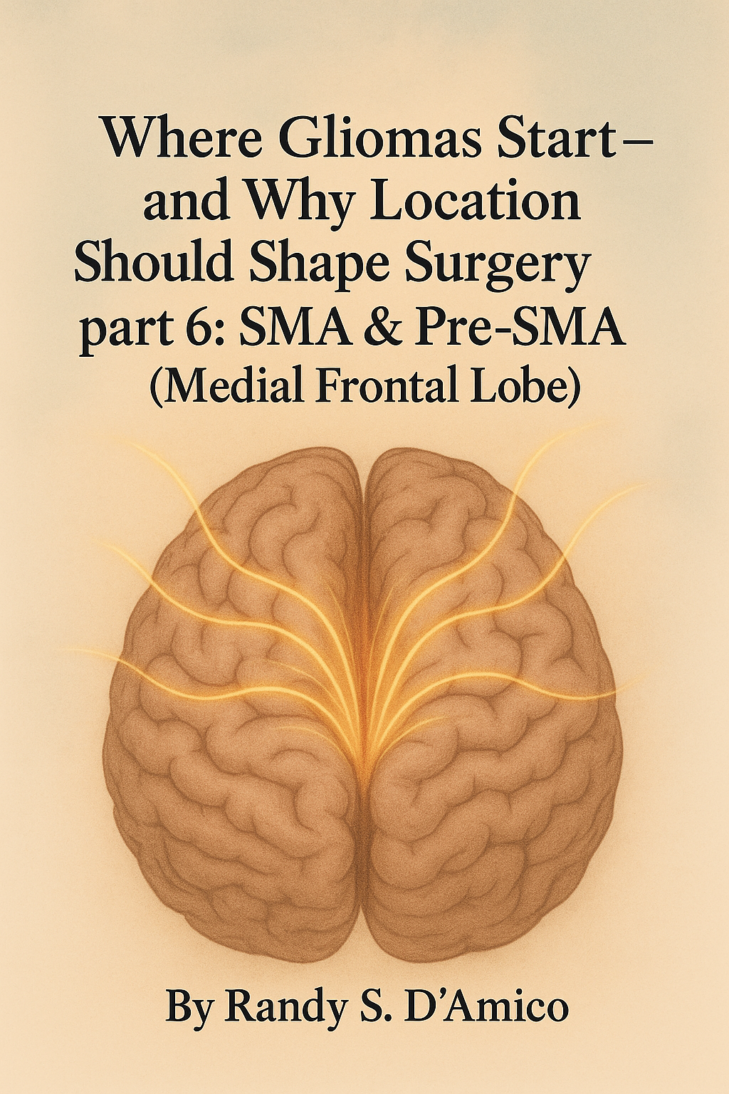
Post 6: SMA & Pre-SMA (Medial Frontal Lobe)
About this series. Plain-English guides to where adult gliomas begin and which brain networks are at risk—so patients and families understand planning, mapping, and recovery.
Why the SMA matters
The supplementary motor area (SMA) and pre-SMA sit along the top-middle of the brain, just in front of the leg part of the motor strip. They don’t drive strength like the primary motor cortex. Instead, they provide the “start signal” for movement and speech, help sequence actions, and coordinate both hands.
Near-SMA surgery often causes a short-term “SMA syndrome”. This involves absence of, or slowed movement/speech (or at its worse paralysis on the other side of the body), and bimanual clumsiness. Fortunately, this usually improves with time and therapy. It’s important to remember that this is not a surgical problem, but the expected physiology of this region. It can be scary though as a patient can awake from surgery with a major deficit. Typically, as long as neuromonitoring of the cortico-spinal tract remains stable, an SMA syndrome can be expected.
What SMA / pre-SMA control (in plain English)
Initiation: getting a movement or a sentence started (not raw power).
Sequencing & switching: finger/face sequences, set-shifting (“now do the other hand”).
Bimanual coordination: both hands working together (zipper, buttons, instruments).
Speech start/fluency: especially pre-SMA for “go signal” and volitional speech.
Common signs you might notice
Slowed start on the body opposite the tumor (e.g., right SMA → left side starts slowly).
Reduced spontaneous speech or brief speech arrest, with intact understanding.
Bimanual clumsiness: tasks with two hands feel off; sequencing breaks down.
Transient weakness can happen, but tone and reflexes look different from pure motor-strip injury.
The nearby “roads” (white-matter)
Short fronto-parietal fibers linking SMA ↔ premotor/motor (execution once the plan starts).
Cingulum / DMN adjacency (motivation, drive, error monitoring) running along the midline.
Callosal interhemispheric fibers (bimanual coupling; the two SMAs talk across the top).
Frontal aslant tract (FAT) anteriorly (speech initiation/volition).
How teams plan surgery (Before • During • After)
Before surgery
Imaging: Map tumor relation to the precentral gyrus (motor) vs SMA/pre-SMA; tractography of FAT, midline cingulum, and callosal fibers.
Baseline testing: finger tapping rate, sequential finger/face tasks, go/no-go, speech initiation (automatic vs volitional).
Expectation-setting: clear discussion that SMA syndrome is common but usually improves over days to weeks (sometimes a few months).
During surgery
Awake (when helpful) or asleep with monitoring:
Tasks: rapid alternating finger sequences, bimanual actions (thumb-index alternation both hands), speech start (counting, days of week, sentence initiation).
Cortical mapping: distinguish SMA/pre-SMA sites (start/sequence disruption) from primary motor (direct strength changes).
Subcortical mapping: respect FAT, midline fibers, and callosal connections.
Strategy: Preserve anterior/medial bridges that support recovery; avoid long retraction on medial cortex.
After surgery
Early rehab: start day 0–1 with external pacing (metronome/timer), chunking tasks, bimanual drills, and automatic → volitional speech progression.
Timeline: greatest improvement in the first 2–6 weeks; plateaus continue to shift over 1–3 months as networks recalibrate.
Return to work/driving/instruments: staged; pair with therapist assessment of initiation and bimanual control rather than strength alone.
Ask your surgeon (patient-facing)
Is my tumor closer to SMA/pre-SMA or the primary motor strip?
What’s my risk of temporary SMA syndrome, and what does recovery usually look like?
Will you monitor the frontal aslant tract and callosal fibers?
Which intraoperative tasks will you use to protect speech start and sequencing?
Bottom line
SMA/pre-SMA gliomas threaten the start, sequence, and two-hand coordination we use all day. When teams plan around the FAT, callosal, and cingulum pathways and use targeted mapping, they can push resection safely—accepting a temporary SMA syndrome to achieve oncologic goals while preserving long-term function.
Fast FAQ
Is SMA syndrome permanent?
Usually no. Most patients improve over weeks, sometimes a few months, with therapy. Persistent, dense deficits point to injury beyond SMA (e.g., primary motor or deep pathways).
Why can strength be normal but movement still “won’t start”?
SMA gives the go signal and sequence plan. Primary motor executes the plan. You can have power without initiation.
Does awake surgery guarantee no SMA syndrome?
No. Awake mapping helps minimize lasting deficits and separate motor strip from SMA. A temporary syndrome can still be expected—and planned for.