Where Gliomas Start — And Why Location Should Shape Surgery
Corpus callosum (“butterfly”) gliomas in plain English—why they cross midline, common symptoms (fatigue, attention, bimanual issues), and how corridor-first care protects function.
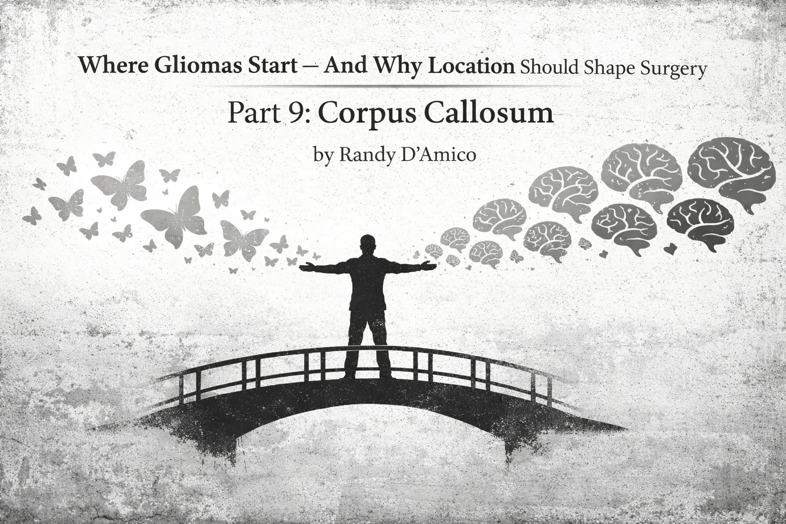
Post 9: Corpus Callosum (“Butterfly” Gliomas)
About this series. Plain-English guides to where adult gliomas begin and which brain networks are at risk—so patients and families understand planning, mapping, and recovery.
Why the corpus callosum matters
The corpus callosum is the brain’s main bridge between the left and right hemispheres. It’s a dense white-matter highway. Tumors that reach it can spread along the bridge, sometimes creating the classic “butterfly” appearance which is really just one tumor mass that extends into both sides.
Takeaway: A callosal tumor is less about a single “lump” and more about a tumor tracking along the brains connections. Treatment decisions focus on function, safety, and realistic goals.
The corpus callosum in plain English
The callosum is organized front to back:
Genu (front): connects frontal lobes → planning, initiation, attention control
Body (middle): connects motor and sensory regions → coordination, both-hand tasks
Splenium (back): connects parietal/occipital regions → visuospatial integration and visual processing
When a glioma involves the callosum, symptoms depend on which segment and which nearby networks are affected.
What dysfunction can look like (real-world examples)
Callosal involvement doesn’t usually produce one dramatic, obvious deficit. It often shows up as changes in integration:
Slowed processing: “Everything takes longer,” mental fatigue spikes quickly.
Split attention issues: harder to multitask; can’t hold two streams of info at once.
Bimanual clumsiness: buttons, tying shoes, cooking with both hands feels off.
Initiation/drive changes: when cingulate/medial frontal networks are involved, this can come across as reduced spontaneity, apathy-like symptoms.
Visuospatial integration issues: especially with posterior callosum involvement - Navigation feels harder, reading/visual scanning gets less fluid.
In some cases, disconnection phenomena can occur (rare, usually more extensive): one hand “doesn’t cooperate,” or actions feel oddly uncoordinated.
Why “butterfly” happens
Glioma cells preferentially migrate along white-matter tracts. The callosum is the biggest tract bundle in the brain. So when a tumor reaches it, it has a ready-made route to the other hemisphere, creating a symmetric, midline-crossing pattern on MRI.
The nearby “do-not-disturb” structures
Callosal tumors sit close to critical midline anatomy:
Pericallosal/callosomarginal arteries (blood supply along the callosal surface)
Cingulum bundle (motivation, attention, error-monitoring—runs just above/around)
Deep ventricular/periventricular pathways (dense fibers near the lateral ventricles)
SMA/medial frontal networks (especially with anterior involvement)
This is why corridor selection is everything.
How teams plan care (Before • During • After)
Before surgery
Define the goal: biopsy for diagnosis? Debulking to relieve mass effect? Maximal safe resection? Minimally invasive Laser Ablation (my favorite…) The answer depends on symptoms, tumor biology, and location within the callosum.
Map networks: relationship to cingulum, SMA, deep periventricular fibers, and frontal/occipital connections.
Baseline function: attention/processing speed, bimanual tasks, initiation and mood/energy profile, plus standard motor/sensory/visual fields.
During surgery
Corridor-first mindset: anterior vs posterior approach depending on genu/body/splenium involvement; avoid vascular injury along the callosal surface.
Monitoring and mapping: motor/sensory monitoring as relevant; subcortical mapping when approaching deep fibers.
A realistic stopping rule: when you’re near pathways where “one more millimeter” risks a disproportionate loss of independence, the safest operation is the one that stops.
After surgery
Rehab focuses on integration: pacing strategies, dual-task training, bimanual coordination drills, and attention scaffolding.
Expect fatigue: cognitive fatigue is common. Recovery plans often emphasize structured routines and graded activity.
Support initiation: if drive is reduced, external prompts and scheduled activation help while networks recalibrate.
Ask your surgeon (patient-facing)
Is the tumor in the genu, body, or splenium, and what functions does that threaten?
What is the plan: biopsy, debulking, or maximal safe resection—and why?
How close are we to the cingulum and SMA/medial networks?
What do you expect to be temporary swelling-related changes vs longer-term risks?
What rehab is planned for attention, fatigue, and bimanual coordination?
Bottom line
Corpus callosum (“butterfly”) gliomas are tumors of connections. The callosum is the brain’s main bridge, so involvement often affects processing speed, attention integration, and bimanual coordination. The best care is goal-driven and corridor-first—protecting midline vessels and nearby medial networks while selecting treatments that preserve independence.
Fast FAQ
Does a “butterfly” pattern mean surgery isn’t possible?
Not automatically. It means decisions must be individualized. Sometimes biopsy plus medical therapy is best; sometimes, biopsy plus LITT plus medicine is best, and sometimes debulking helps symptoms. The key is a realistic goal and a safety-first plan.
Why do people feel “foggy” or slow with callosal tumors?
Because the hemispheres share work through the callosum. When the bridge is disrupted, integration becomes inefficient.
Can rehab help?
Yes—especially with cognitive pacing, attention strategies, and bimanual retraining. Even when MRI changes are limited, training can improve day-to-day function.